Interpretation of equine vision is complicated and highly subjective. Understanding how your horse sees the world, and being able to identify when vision is compromised, will enable you to work with your veterinarian to manage the health of your horse’s eyes.

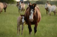
Paint horse looking at observer.
Many equine ophthalmological procedures can be performed on standing, sedated horses thereby reducing the risk of complications from general anesthesia.
© 2016 by Kent Weakley
We collaborated with the UC Davis Ophthalmology Service to draw attention to some things you might not know about equine ophthalmology.
1. An equine eye issue is often urgent. When it comes to your horse's eyes, it is best not to take any chances. If you notice an eye problem, contact your veterinarian right away. Do not administer any medications without talking to your veterinarian. Using the wrong medications can result in serious complications.
2. Horse eyes are among the largest of all land mammals. Only whales, seals, and ostriches have larger eyes. Horses' large eyes, with large corneas, allow a significant amount of light to enter. Their pupils can dilate to an area three times larger than a cat or dog and six times that of a human. Since horses are prey animals, their large eyes enable them to detect even slight motion, including that plastic bag blowing outside of the arena!

3. Horses have both monocular and binocular vision. Horses' eyes are located on the sides of their heads, providing them with extensive monocular vision that allows for a panoramic view of up to 350 degrees - almost a complete circle. Horses raise their heads to enable binocular vision with both eyes ahead of them, with a visual field of 55-80 degrees.
4. Horses have small blind spots in their field of vision. These are about the width of the horse's body and include areas above and perpendicular to the forehead, directly below the nose, and directly behind the horse. This is important to remember when working around horses on the ground, as well as when riding.
5. Horses have better vision in low light conditions than humans, but not quite as good as cats. Horses have a reflective tapetum lucidum, a structure that lies behind the retina and increases the light available to photoreceptors, enabling them to see in dim light. It has a slightly different structure from the tapetum of carnivores, like cats, which are highly reflective. Human eyes lack a tapetum altogether. Studies have shown that horses can readily discriminate between different shapes and successfully navigate in a room with such low light levels that humans could not see at all!

6. The menace response is learned; foals develop it at one to two weeks of age. The menace response is a protective blink reflex, sometimes accompanied by movement of the head and/or neck, to avoid a threat. It is assessed by moving an object towards the horse's eye. It is important, however, to avoid touching the eyelashes or creating a significant current of air, which could interfere with the results. A decreased menace response, or lack of a response, can indicate an issue with vision anywhere from the eye all the way to the cerebral cortex of the brain.
7. Pioneer 100 Horse Health Project horses underwent ophthalmologic examinations and electroretinograms (ERGs). The “Pioneer 100 Horse Health Project”, a first-of-its-kind precision medicine study in horses, included evaluations by the UC Davis Ophthalmology Service. Results from these examinations showed lipofuscin deposits in some horses, which are associated with aging in horses affected with equine motor neuron disease (EMND).

8. Horses' prominent eyes are prone to injury from foreign bodies and corneal ulcers. In many cases, foreign bodies occur in the form of plant matter, such as a piece of hay, cactus spine, or seed awn. Foreign bodies in the eyes can cause corneal ulcers (ulcerative keratitis), lacerations, and infections, which can result in serious trauma and loss of vision.
9. Bacteria and fungi can cause eye infections. Both thrive in the environment and can enter horses' eyes through cuts and scratches (even small ones). Infections can occur on the surface or deeper into the eye. Fungal infections often cause painful ulcers and abscesses that can result in vision loss if not properly treated. The specific type of fungus involved, which is influenced by geographic location and environmental factors, can affect disease severity, but ALL fungal eye infections are serious.
10. Many equine ophthalmological procedures can be performed on standing, sedated horses. This reduces the need
for many horses to undergo general anesthesia, opening up procedures such as cyclosporine implants, enucleations, and corneal-conjunctival grafts to horses that otherwise are at risk of complications from general anesthesia. It also reduces costs. The UC Davis Ophthalmology Service works closely with the UC Davis Anesthesia Service to offer these procedures for eligible patients.
Article by Amy Young - UC Davis Horse Report - School of Veterinary Medicine
