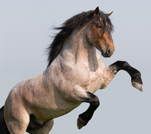
The Orthopaedic Research Center at Colorado State University is known worldwide for joint problem prevention and healing research in the horse with some recently expanded work in human athletes.

Research benefitting horses and humans
Proceeds from annual Stallion Auction at CSU support orthopaedic research to find new methods to rehabilitate damaged joints, prevent or decrease joint disease and musculoskeletal injuries.
The 14th Annual CSU Stallion Auction to benefit equine research at Colorado State University will be held January 9-12, 2013. Osteoarthritis remains a common and debilitating disease in humans, horses and other mammalian species, despite advances in diagnosis and treatment.
The ORC uses state-of-the-art research to find new methods to rehabilitate damaged joints, prevent or decrease joint disease and musculoskeletal injuries, offer early detection, and develop better treatments that prevent permanent joint damage for both horses and humans.
Proceeds from the annual auction support continued equine research into a multitude of orthopaedic issues that benefit horses and humans alike.
For example in one case file, a 6-year-old Quarter Horse mare was presented to the Colorado State University Veterinary Teaching Hospital for right fore lameness of three months duration. The mare had recently undergone a change of ownership, and had a corresponding increase in exercise as a ranch horse and barrel racer.
On lameness evaluation, the mare was 3/5 lame on the right fore and was moderately positive to flexion of the distal limb. Perineural anesthesia was performed, which showed 60 percent improvement when 1.5 ml of the anesthetic was placed around the medial and lateral palmar digital nerves.
Additional improvement was seen with perineural anesthesia at the medial and lateral palmar digital nerves at the level of the base of the sesamoids. The following day, an intra-articular block was performed in the distal interphalangeal joint which improved the lameness by 60 percent.
Radiographs of the distal limb were taken including four views of the coffin joint. The lateromedial radiograph revealed several mineral opacities at the palmar aspect of the distal interphalangeal joint (Fig. 1). Ultrasound examination showed that the mineral was at the palmar lateral aspect of the joint and impinging or possibly located in the collateral suspensory ligament of the navicular bone.
An MRI was recommended to more accurately determine the location of the calcifications. The MRI revealed significant osteochondral fragmentation and was was the key in making the correct diagnosis.
A large cartilage-covered fragment was found at the lateral aspect of the palmar coffin joint. The fragment was approximately 2-cm long and 1-cm wide, and completely covered in cartilage with another smaller fragment next to it.
A corresponding defect was seen at the lateral palmar aspect of the second phalanx. This defect was covered in some areas with fibrocartilage. The fragments were removed in several pieces due to the constraints of the size of the surgical instruments, and to maintain distension of the joint. Some soft, irregular bone was removed from the defect in the distal phalanx using a curette. The joint was carefully inspected to ensure removal of all fragments and then thoroughly lavaged.
Post-Operative Care
The mare was kept at CSU overnight for treatment and monitoring. She was then sent home with instructions to stall rest for two weeks, followed by four weeks of hand-walking. At six weeks post-surgery, the mare returned for a recheck exam. Her lameness was much improved (1/5 lame on the RF and only detectable on a small circle) being mildly intermittently lame in a right circle on hard ground. She had mild sole pain likely due to not being shod.
The mare was shod and gradually returned to normal work.
Case Summary
We are fortunate to have several imaging modalities including MRI, which allows us to localize the lesions within the hoof capsule and determine severity. Although our MRI does require general anesthesia for use, the images acquired are some of the most detailed available. It was through the use of the MRI that we were able to determine that the fragments were not just mineralization of the soft tissues (collateral suspensory ligament of the navicular bone), as this was difficult to determine with radiographs and ultrasound.
The prognosis and treatment for mineralization of the soft tissues is much different than for joint fragments, and therefore MRI was paramount in diagnosing and treating this case.
Imaging was especially important in this case as a developmental lesion in bone maturation causing a free fragment of cartilage-covered bone (osteochondritis dissecans) has not been documented previously in the palmar aspect of the distal interphalangeal joint.
As the bone fragment was covered in cartilage on all sides, it is likely that this fragment was not traumatic in origin, but developmental. There is no previous publication on osteochondritis dissecans in the palmar distal interphalangeal joint, and this may be the first of its kind.
Clinicians involved: Dr. Lacy Kamm, Dr. Laurie Goodrich, Dr. Myra Barrett, and Dr. Natasha Werpy
Information from Department of Clinical Sciences, College of Veterinary Medicine and Biomedical Sciences, Colorado State University
