The Equine Cancer Society is a non-profit organization dedicated to the education of all horse owners on equine cancer. ECS also helps to raise funds for equine cancer research. In their effort to educate horse owners, leaders in the Society offer clinics at convenient places.

Sarcoid tumors on horse's skin
The Equine Cancer Society is a non-profit organization dedicated to the education of all horse owners on equine cancer and also raises funds for equine cancer research.
© 2012 by Malcolm Morley
As most people know, cancer is a state of cellular growth occurring when normal cells become abnormal, and continue to grow abnormally, characterized by the ability of those abnormal cells to subdivide and grow while limiting the ability of normal cells to do the same.
These cancerous cells may invade healthy tissue as a more diffuse blanket of cells throughout the tissue similar to roots of a tree spreading throughout the ground. The tumors themselves release chemicals into the body that cause several different side effects such as weight loss, depression, anorexia, fever, anemia just to name a few.
Once a cell type or tissue becomes cancerous it can spread through the blood or lymph system to all regions of the body and become tumors anywhere.
Common Cancers Found in Horses:
Melanoma
This type of skin cancer can occur in any dark-skinned horse, but is most common in gray horses. Melanomas are tumors originating from the cells that produce skin pigment (melanin). A high percentage of gray horses over 15 years of age develop melanomas. They may appear any time after age 4 or 5, and growths that appear on a relatively young horse are more likely to become malignant more quickly than those that appear on older horses. Lumps may appear singly or in groups.
Squamous Cell Carcinoma
This type of skin cancer is usually quite visible and readily detected. It often appears around the anus or genitalia. It also occurs around the eyes or eyelids of the horse.
It is most common in areas of unpigmented skin, especially those that have little hair covering—such as under the tail, around the mouth, or on the sheath. Cancer of the eyelid is fairly common in Appaloosas, Paints, Pintos or any horses with white faces, large white markings with pink skin around the eye or any horse with light skin. Tumors surrounding the eyeball are fairly common in sunny regions; intense sunlight irritates the eyes and encourages growth of this type of cancer. Unpigmented skin has little protection against the harmful effects of the ultraviolet rays.
Cancer of the third eyelid can spread to the surrounding tissues and may necessitate removal of the eye to save the horse. If caught early, the removal of the small tumor can prevent eye removal and long term resolution.
Horse owners should try to be aware of any changes around the eyes. The growths often occur on the third eyelid or on the lower eyelid’s inner surface. They may appear as a single raised bump or a raw surface that looks like a runny sore. A bump or red area is not always cancer, but if it becomes larger, redder or more irritated, the horse should have immediate veterinary attention.
This type of cancer will continue to grow and can spread to nearby tissues, eventually killing the horse unless removed.
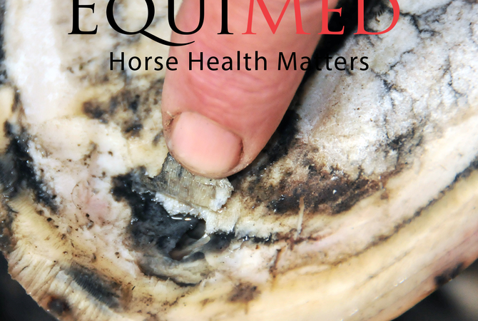
Sarcoid Tumors
Cancer can be defined as an uncontrolled proliferation of cells, and this certainly describes a sarcoid. The good thing about sarcoids is that they don’t usually spread internally; they don’t travel through the bloodstream to other parts of the body. They stay in their local area, even if they get huge. If not removed early, however, the tumor may become so large that it disables the horse.
Sarcoids are one of the most common skin tumors of horses. They are believed to be caused by the bovine papilloma virus. They can be treated with chemotheropy, such as cisplatin, or removed surgically. Unfortunately, sarcoids are known to return if not all of the effected tissue is removed.
Lymphosarcoma
This is the desease of the lymph tissue. The lymph tissue becomes cancerous, often starting with one lymph node and spreading to others. The lymphatic system is the body’s defense network and lymph nodes are small nodules throughout the body that filter lymph, destroy infections, and produce white blood cells. People often refer to these nodes as “glands.” Along with the lymph nodes, there is also lymph tissue that is diffusely spread throughout organs such as the intestines and the skin. Any one of these “glands” or lymph tissue can become cancers, hence the symptoms of the disease can vary greatly depending on the location of the cancerous tissue. There are four general types of lymphosarcomas which are categorized on location. The four main types are generalized (multi-centric), intestinal, mediastinal and cutaneous.
The generalized form is the most common and includes multiple peripheral and internal lymph nodes. Basically, it is tumors through the lymph nodes of the body. Common sites are the nodes (glands) around the throatlatch, in between the jaw, at the base of the neck, superficial inguinal, mesenteric, and the pectoral region. The most common clinical sign of this type of lymphosarcoma is large masses on the chest, at the base of the neck, under the jaw, and at the throatlatch. Ventral edema is very common as well as weight loss. Sometimes the diffuse lymph tissue within the skin can also be affected which manifests itself as a severe ulcerative dermatitis, where the skin literally erupts with crusting sores that don’t heal. This form also has the lowest survival rates, often only weeks to a few months.
Intestinal lymphoma involves the diffuse tissue within the intestinal wall. Involvement in this area causes malabsorption problems of the intestines. This leads to severe weight loss, diarrhea and sometimes colic. Mediastinal refers to the lymph nodes within the chest in between the lungs. Tumors in this region can cause coughing, increased heart rates, fluid on the chest and even fluid within the chest.
The best and least deadly form of this disease is the cutaneous form which, is lymph nodes that turn into tumors under the skin and don’t migrate throughout the body. Horses with these types of tumors typically do very well and live fairly long with minor clinical symptoms.
Unfortunately, if large tumors are not visible, diagnosis can be difficult. There is also overlap of the different types, which leads to further diagnostic challenges. A biopsy of the tumor can confirm the diagnosis, but can sometimes introduce tumor cells into the blood stream which could cause the cancer to spread further. An ultrasound of the tumor is typically very characteristic and diagnostic. Often a diagnosis is made from clinical signs such as weight loss, masses, depression, edema of belly and legs, fever, diarrhea, skin crusting and scaling, coughing, high heart rate, blood changes such as anemia and a variety of other symptoms caused by the tumors that affect the tissue they are close to.
Breast and Ovarian Cancer in Mares
Breast Cancer, or more correctly, mammary neoplasia, in mares is uncommon in comparison to rates among women. Abbatior studies (cited by Equine Disease Quarterly July 2008) report an incidence of barely 2%, and less than a dozen published cases known to exist. But of that compilation, all but one were malignant. And the results of necropsies, conducted between 1994 and 2008 by Livestock Disease Center at the University of Kentucky on 11 mares diagnosed with malignant mammary neoplasia (cancer), have also concluded that equine mammary cancer is "much more likely" to be malignant the benign and carries a "poor prognosis" for long term survival.
Prostate and Testicular Cancer in Stallions, Geldings
Tumors of the testicles are uncommon, the incidence is unknown as most male horses are castrated at an early age, but the tumors are unilateral. Testicular tumors are divided for descriptive purposes into either Germ Cell Tumors or Non-germinal tumors. A Seminoma is the most popular of the germ cell tumors. This type usually effects the aging stallion. A Teratoma is another germ cell tumor. These tumors are usually oval or round in shape and are most common in the abdominal testicles. They are rarely malignant. Non-germinal tumors are rare in horses, but can cause increased testosterone production,
Prostate glands in male horses are susceptible to the same cancers, conditions and issues as human, with not-so-rare cases of death per Dr. Kuehnle. This type of cancer is becoming more of a problem as horses are exposed to abnormal internal, and external factors.
As the Equine Cancer Society points out: The important thing to remember is if you see any change in your horse, his routine, his demeanor, unexplained physical changes such as lumps or sores that do not heal, seek immediate veterinary care. Please remember a horse is a prey animal, and as with any prey animal, signs of weakness or unsoundness attract predators, therefor a horse's survival instinct will cause it to hide signs of weakness.