According to David Bolin, DVM, PhD of the Veterinary Diagnostic Laboratory at University of Kentucky, congenital cardiovascular malformations are rare in horses with an estimated prevalence of 0.1-0.5%.

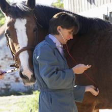
Veterinarian checking horse's heart sounds
Although congenital cardiovascular malformations are rare in horses, typical clinical signs can include stunted growth, exercise intolerance, heart murmur, tachycardia, respiratory distress, and cyanosis(a bluish tinge on mucous membranes).
Male and female horses are similarly affected, and a clear-cut breed predilection is not evident. Cardiovascular malformations are broadly classified as either simple (a single anomaly) or complex (multiple coexisting anomalies).
Each major category is further subdivided on the basis of the tissue affected: myocardium (heart muscle), blood vessels, or valves. Complex malformations typically involve multiple tissues and have the least favorable prognoses.
Clinical signs vary in severity and the age of onset. Typical clinical signs can include stunted growth, exercise intolerance, heart murmur, tachycardia, respiratory distress, and cyanosis.
Affected horses are frequently found dead, however not all horses with cardiovascular malformations display clinical signs or die from the anomaly. In some cases, the cardiovascular defect is only identified at necropsy as an incidental finding and is not related to the cause of death.
Ventricular septal defect (VSD) is the most frequently reported congenital cardiac anomaly in the horse. This anomaly is represented by a patent channel in the interventricular septum that allows communication between the two ventricles, which play a critical role in pumping blood.
The channel can result in altered pressures within the heart, the shunting of blood through the channel, compensatory hypertrophy (enlargement of the heart muscle), and systemic abnormalities (e.g. cyanosis) in severe cases.
Horses with small defects may be asymptomatic or develop clinical signs at a later age. VSDs are also components of complex cardiac anomalies such as Tetralogy of Fallot and truncus arteriosus.
Other defects described in the horse include:
- Abnormal communications between the atria. Examples: atrial septal defect and patent foramen ovale.
- Abnormal communication between the great vessels. Example: patent ductus arteriosus.
- Malformed great vessels. Example: common truncus arteriosus (the division between the aorta and pulmonary artery does not develop and a single vessel leaves the heart).
- Abnormally positioned great vessels. Examples: complete transposition (the right ventricle pumps blood into the aorta and the left ventricle pumps blood into the pulmonary trunk) and double-outlet right ventricle (both the aorta and pulmonary trunk arise from the right ventricle).
- Heart valve abnormalities. Example: tricuspid valve atresia.
- Tetralogy of Fallot, which consists of a dextra-positon of the aorta, pulmonic stenosis, ventricular septal defect, and right ventricular hypertrophy.
Archives at the University of Kentucky Veterinary Diagnostic Laboratory were searched from 2010 to 2015 for cases of congenital cardiovascular malformations in the horse.
Over that period, 18 cases were identified. Fourteen were in Thoroughbreds, two in American Saddlebreds, and one each in the Arabian and Standardbred breeds. Twelve of the animals were female and five were male; the sex of one animal was not identified.
Ten cases of ventricular septal defect, which included nine simple and one complex anomalies, were diagnosed. The complex anomaly was associated with pulmonic stenosis.
Two cases each of Tetralogy of Fallot and truncus arteriosus with VSD were identified, and single cases of atrial septal defect, over-riding aorta, right ventricular hypoplasia with pulmonary atresia and moderator band dysplasia were reported.
Article by David Bolin, DVM, PhD
Veterinary Diagnostic Laboratory
University of Kentucky
