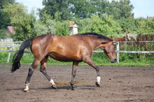
As part of the Piper Clinic Trainers Series in April 2013, at the University of Minnesota, Drs. Nicolas Ernst and Kyla Awes shared a presentation about back pain from the perspectives of an orthopedic surgeon and a chiropractor working together.

Gait as an indication of horse back pain
When diagnosing back pain in horses, attention is directed at determining whether pain arises primarily from malfunction of bones and muscles in the back or whether it is secondary to malfunction of structures elsewhere.
Ernst emphasized the importance of a specific diagnosis for the cause of back pain and Awes discussed how the way a horse is trained to use its body affects the back and the importance of a team approach to successful management of a horse with back pain.
Attention is initially directed at determining whether pain arises primarily from malfunction of structures in the back itself or whether pain in the back is secondary to malfunction of structures elsewhere in the body.
Secondary back pain is far more common than primary back pain. Secondary back pain occurs when a horse carries itself differently, which often happens when there is soreness in one of the limbs.
Hindlimb or forelimb lameness alters the way the back functions, and this additional strain results in back pain. (Think about wearing high heels all day!) It is therefore imperative that a thorough and complete lameness evaluation be performed in horses with back pain.
Primary versus secondary back pain in horses
Determining that back pain exists is relatively straightforward. The more difficult question is what is causing back pain? An accurate diagnosis is necessary to determine the future use of the horse (prognosis) and select the correct therapy.
Secondary back pain can also result from other disorders that affect the way a horse carries itself: neck problems, sacroiliac disease, pelvic fracture, lack of fi tness or supporting back musculature, muscle diseases, dental problems, rider or horse’s inability to perform at desired level, temperament, and improper fi t of tack. In cases of secondary back pain, once the source of the problem is identifi ed and treated, the horse can gradually be put back to work in the right frame and back pain diminishes.
Primary back pain can result from soft tissue injuries to the ligaments, tendons, and muscles supporting the spine or from boney injuries to the vertebrae (e.g. kissing spine). Nerve compression is also possible. Each of these causes of primary and secondary back pain requires a different treatment regimen.
Five of the most common causes of back pain in horses are:
1. "Kissing Spine"
There is a great degree of controversy surrounding the diagnosis of overriding/ impingement of dorsal spinous processes, also known as "kissing spine."
Impingement occurs when dorsal spinous processes (upward bone projections off of each vertebrae) that are next to each other rub together. Pain is caused by repetitive, traumatic contact between neighboring dorsal processes or from a primary injury to the supporting soft-tissue structures of these vertebrae.
The clinical signs associated with this disease may include reduced mobility of the spine, limited side-to-side back mobility due to muscle spasms, and painful or violent responses when the saddle is placed on the horse or when the rider mounts.
These painful or violent responses associated with saddling or mounting should be differentiated from improper saddle fit. The controversy surrounding kissing spine exists because determining whether vertebrae that touch are actually causing pain is very difficult. Radiography (X-rays) and nuclear scintigraphy (bone scan) can assist in a diagnosis; however, these diagnostic techniques are not the final answer in the diagnosis of kissing spine.
If a diagnosis of kissing spine is highly likely, a combination treatment protocol may include rest, non-steroidal antiinfl ammatory medications, local injections of anti-inflammatory agents, acupuncture for pain, physiotherapy, or, in severe cases, surgery. Prognosis depends on severity and whether bone, supporting soft tissue, or both are involved.
2. Supraspinous ligament injury
Supraspinous ligament injuries usually appear as a lump on the horse's back and can be associated with pain and thickening of the area under the skin over the spine. These injuries often occur in the region of the back where the saddle sits.
Diagnosis can be made using ultrasonography. Treatment usually includes rest to allow the ligament time to heal, non-steroidal anti-infl ammatory medications, and rehabilitation of controlled stretching and a gradual increase in exercise. Prognosis and timeline to recovery (usually two to six months) depends on the extent of injury.
3. Vertebral facet joint arthritis
Each vertebrae of the spine has four articular facets that form the joints between neighboring vertebrae. Both trauma to the spine and everyday wear and tear can lead to vertebral facet joint arthritis. Injuries and trauma to the spine, which can occur with a fall, can result in unstable vertebral joints. The musculature surrounding and supporting the spine must compensate to stabilize the spine, which produces painful muscular spasms.
With severe arthritis of the vertebral facets, adjacent vertebrae may fuse, leading to decreased back mobility. Clinical signs seen in horses with vertebral facet joint arthritis may include lameness, abnormal gait, a stiff-looking or short-strided trot, difficult downward gait transitions (e.g. canter to trot), and reluctance going downhill or jumping.
Diagnosis is made using a combination of radiography, ultrasonography, and/or a bone scan.
Successful treatment of vertebral facet joint arthritis involves rehabilitation and development of proper muscle support for the spine. Other treatment options that may be used in addition to rehabilitation include non steroidal anti-inflammatory medications, ultrasound-guided vertebral facet injections, methocarbamol, and rest.
Prognosis depends on a number of factors, including the extent and severity of the arthritis as well as the intended use of the horse. Dressage horses tend to be more successful in return to work than jumpers or three-day eventers since these horses require different types of movement in their spine.
4. Spondylosis
Spondylosis is a degenerative condition affecting the vertebrae. It usually affects older horses.
It is caused by mechanical stress on the area, which results in reduced mobility of the spine and, in the end stages of disease, complete fusion of the vertebrae.
Clinical signs include generalized stiffness and reluctance to work. Spondylosis occurs more commonly in eventers and jumpers and also in working draft horses that endure large amounts of force on their backs while carrying heavy loads.
Treatment relies on non-steroidal antiinflammatory medications alone; there is no surgical technique that can stabilize the spine of these horses. Prognosis for an athletic career is guarded, because there is no way to reverse the disease process and fusion of vertebrae.
5. Sacroiliac disease
Sacroiliac disease affects one or more of the bones, ligaments, muscles, and joints (sacroiliac and lumbosacral joints) of the sacroiliac region. There are generally three types of injury in the sacroiliac region that can lead to sacroiliac disease. The most common cause of sacroiliac disease is damage to soft tissue and ligaments; this can occur from muscle weakness, fatigue from improper training, overuse, or repetitive stress injuries. Profound trauma of the ligaments, such as with a fall, can also lead to sacroiliac disease. The least common cause of sacroiliac disease is sacroiliac joint problems.
Clinical signs of sacroiliac disease may include shortened stride length in one or both hind limbs, asymmetry of the hind end, difficulty in downward gait transitions, or reluctance to go downhill. Signs are usually best seen during the canter.
A lameness exam and local anesthetic blocks might help localize pain to the sacroiliac area. The imaging modality that can give you the best indication of degeneration or damage to the sacroiliac region is a bone scan, because ultrasound and radiographs are often not able to penetrate the necessary structures.
Treatment of sacroiliac disease often involves stall rest, non-steroidal antiinflammatory medications, and intensive rehabilitation to rebuild musculature.
Prognosis depends on the cause, extent,and severity of injury. When putting horses back in work that have had back soreness, it is essential that they work with proper posture and carriage. The way a horse moves and uses its body has a direct impact on its back. A horse can initiate movement in different ways that are more or less effi cient and put more or less strain on the back.
For example, if a horse uses its neck or head and "falls on the forehand," it isn't utilizing the powerful hindquarter muscles as much. This type of motion is inefficient and fails to build core strength critical for a healthy back. If a horse is engaged and uses its hindquarters to initiate motion, it is moving in a more effi cient manner.
Engagement of the hindquarter muscles in motion and the resulting efficient movement is important because it means that the horse is conserving energy, building core strength, and reducing wear and tear on the body. This is even more important when we add the weight of a saddle and rider onto the horse's back. The horse must be trained to carry itself and the rider in a way that lifts its back against this weight.
Training young horses in a "long and low" position facilitates the lifting of the back, which makes it easier for the horse to active their core muscles. A neck that stretches forward and down, while avoiding curling the nose inwards, adds further stretch to the back. Training a horse in this way builds its neck, abdominals, and sublumbar core muscles, all of which are responsible for lifting the back and stabilizing the spine.
Horses ridden with a high head carriage and hollowed back or horses with a long and low head carriage but with a braced back are not activating their core muscles or lifting their back. The reason the horse's core muscles must be strengthened, like when training in a long and low position, is because these muscles must stabilize the pelvis and spine sufficiently to transmit the forces generated by the powerful hindquarter muscles during engagement.
