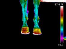
Since the late 1990's use of thermography has increased in both finding and preventing causes of lameness. Originally used mainly with race horses and show animals, current usage has expanded in many veterinary practices.

Thermal photo showing heat in hooves
Thermography measures the presence of heat which is one of the cardinal signs of inflammation resulting from hemorrhage, trauma, tendon or joint infection in the horse.
© 2013 by Miriam Rieck
Heat or inflammation is generally a result from hemorrhage, external or internal trauma, tendon or joint inflammation and/or infection
Thermography is a non-invasive technique, whereby a camera with a sensor measures the infra-red emissions from a horseâs body. Once the information is recorded, it enters the camera and the temperature variations are depicted in the display with different colors corresponding to different temperatures.
Essentially the sensors see into the horseâs body,enabling the veterinarian to compare one leg or one site with another. After images are recorded in the camera they are then downloaded into a computer that analyzes the data and generates a report which indicates the relevance of the temperature variations.
The reason that heat detection is important is because the presence of heat is considered one of the cardinal signs of inflammation. The human hand is able to appreciate, in some circumstances, a change in temperature of 1.0 degrees C, while this camera can detect changes down to 0.1 degrees C, a ten-fold improvement. In addition to heat, the camera will also appreciate cold areas.
Heat or inflammation is generally a result from hemorrhage, external or internal trauma, tendon or joint inflammation and/or infection. Conversely, cold may be seen with decreased circulation, non-active swelling such as edema or in the presence of obstruction of blood flow.
To evaluate the use of thermography in equine medicine, a three-phase study was conducted. In the first phase, six horses were examined thermographically, before and after exercise, to determine a normal thermal pattern.
In the second phase, nine horses with acute and chronic inflammatory processes were examined thermographically. In the third phase, thermography was used to evaluate the effectiveness of anti-inflammatory drugs on chemically induced inflammatory reactions. All normal horses tested had similar infrared emission patterns.
A high degree of symmetry between right and left and between front (dorsal) to rear (palmar, plantar) in the legs distal to the carpus and the tarsus existed in the tests. The warmer areas of the thermogram tended to follow major vascular structures. The coronary band was the warmest area of the leg.
Heat increase due to exercise did not substantially alter the normal thermographic pattern. Use of thermography in clinical cases successfully detected a subluxation of the third lumbar vertebra, a subsolar abscess, alveolar periostitis and abscess, laminitis, serous arthritis of the femoropatellar joint, and tendonitis.
Thermography was effective in quantitative and qualitative evaluation of anti-inflammatory compounds in the treatment of chemically induced inflammation.
One of the main advantages of equine thermography is the detection of subclinical inflammation. A recent study was undertaken to determine a specific threshold value of temperature change indicative of subclinical inflammation of the lower parts of the horse's limb.
The study involved monitoring 20 racehorses over a period of 10 months. Temperatures of the third metacarpal region were measured every 3 weeks, allowing the average temperature differences to be ascertained between the same areas of forelimbs from the dorsal and palmar aspects in each session.
Additionally, ultrasonographic and radiographic standard examinations of lower part of forelimbs were conducted to diagnose any pathological conditions of lower forelimbs. To determine the threshold value of temperature difference the receiver operating characteristic curve method was used, based on thermographic examinations of the same measured area in 20 horses.The threshold value of temperature difference indicative of subclinical inflammation was found to be 1.25°C.
In conclusion, research has determined that thermography can be used as a quick and practical diagnostic tool of subclinical inflammation. These results provide additional support for the continued study of the use of thermography in diagnosing and treating equines.
