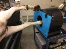
The UC Davis veterinary hospital recently acquired a positron emission tomography (PET) scanner, becoming the first veterinary facility in the world to utilize the imaging technology for equine patients. In association with the UC Davis School of Veterinary Medicineâs Center for Equine Health (CEH), the hospital will launch use of the PET scanner in the summer of 2016. The unit has been acquired for research and clinical studies on lameness diagnosis in horses.

Using PET imaging scanner on horse's leg
The UC Davis veterinary hospital recently acquired a positron emission tomography (PET) scanner, becoming the first veterinary facility in the world to utilize the imaging technology for equine patients.
© 2016 by UC Davis
While most other imaging techniques provide âmorphologicalâ information (identifying changes in size, shape or density of structures), PET is a âfunctionalâ imaging technique, observing activity at the molecular level â detecting changes in the tissue before the size or shape is modified. Once morphological changes have occurred, PET can tell whether the changes are still active or not.
âIn practicality, that means two things,â said Dr. Mathieu Spriet, a UC Davis veterinary radiologist. âOne, PET can detect lesions that other advanced modalities do not identify, and two, it can tell us if a lesionâidentified with another modalityâis a significant injury or not.â
The equine PET scanner has produced initial dataâobtained last year at UC Davis during a research project using a prototype of the new scannerâthat demonstrates great success for bone imaging. The project revealed several PET capabilities for equine imaging:
- identified small areas of bone remodeling at the attachment of tendons or ligaments missed with other modalities
- showed increased activity in bone adjacent to joints, where degenerative changes are known to occur, before morphological changes were present
- revealed increased activity in some joint fragments whereas other joint fragments appeared quiet
- demonstrated that some areas of bone proliferation were active, whereas others were quiescent
âPreliminary data suggests that PET will be the next big revolution in equine imaging since the development of MRI,â said Spriet.
In order to confirm these findings and further define the role of PET in lameness imaging, UC Davis will launch a clinical trial in the fall of 2016. Horses likely to benefit from enrollment in the trial are:
- horses for which other advanced imaging modalities (MRI, CT or nuclear scintigraphy) have failed to identify the cause of the lameness
- horses for which the results of other imaging modalities are confusing due to the presence of multiple abnormalities or equivocal findings
PET has also shown great promises in evaluating soft tissue lesions, in particular regarding laminitis and tendon lesions. Research studies gathering further information in these specific areas will commence shortly at UC Davis. As more data becomes available, additional clinical trials will likely develop.
Support for research projects and clinical trials involving PET, as well as the acquisition of the scanner, was provided by the Grayson-Jockey Club Research Foundation and private donations through CEH.
âWeâre grateful that our donors can see the vision of what these new technologies can bring to equine health,â said Dr. Claudia Sonder, director of CEH. âWe look forward to this PET research translating to cutting-edge clinical applications at the UC Davis veterinary hospital.â
To learn more about PET at UC Davis, please see here.
About the Veterinary Medical Teaching Hospital
The William R. Pritchard Veterinary Medical Teaching Hospital at the University of California, Davisâa unit of the #1 world ranked School of Veterinary Medicineâprovides state-of-the-art clinical care while serving as the primary clinical teaching experience for DVM students and post graduate veterinarian residents. The VMTH treats more than 51,000 animals a year, ranging from cats and dogs to horses, cows and exotic species. To learn more about the VMTH, please go to www.vetmed.ucdavis.edu/vmth
