Horse owner observations of lameness
So, your horse is limping! What can you do to address the problem and how should you go about it? It is in your best interest as a horse owner, to take prompt action any time you have the least suspicion that something is not right with your horse, especially when it comes to any indications of lameness.
Careful observation is the key to identifying what is causing the lameness. When your horse is lame, it is important to follow up with any treatment your farrier or veterinarian recommends. By using your eyes and your hands as you examine your horse and observe your horse in action, you may be able to locate the cause of the lameness.
Most lameness problems involve a structure in or below the knee or hock, so, as you move forward with your observations, pay close attention to the legs and feet of your horse.
1. Begin with the feet, since many cases of lameness begin here.
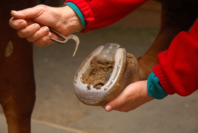
- Pick the horse's feet and make sure no rocks are wedged into crevices.
- Look for dark spots that might indicate a bruised sole. Check for discharge or odor.
- Look for cracks, and check to see if the hooves have been trimmed too short, if a nail is close to the sensitive structures of the hoof, or if the shoe doesn't fit properly.
- Move the heels, tap the hoof wall, and use hoof testers if you have them.
- Feel the hooves. Is one hoof warmer than the others? Do you find a pounding pulse? If so, these are indications of injury or possible abscesses.
2. Check the lower legs for heat and swelling that might indicate inflammation.
- The horse may have an injured tendon or ligament.
- Note any abnormal stance such as favoring one leg, pointing the toe, or a dropped fetlock.
- Look for wounds or injuries to the lower legs.
3. Check the joints carefully for heat and swelling
- Horses can suffer from arthritis and other degenerative diseases
- Has the stifle slipped and locked?
- A horse may have a bone chip floating in the joint.
- Flex and extend the joint to observe range of motion and to check for pain.
- Note any areas that are inflamed, as evidenced by heat and swelling.
4. Check the neck and back for symmetry, posture, and contour.
- As you move your hands over the horse's neck and back, notice any indications of swelling, pain, heat, inflammation, or loss of muscle tone.
- Does your horse flinch when you approach with the saddle or move away from your touch when you attempt to brush the neck or back areas?
- Are there any changes in range of motion?
5. Now, check the horse's gait on a level, even surface with several maneuvers including a walk and a trot, in a straight line and in a circle.
- As you observe from the side, rear, and front, try the horse on soft and hard surfaces, as well as up and down inclines.
- Note any abnormal head movement, including a bobbing of the head as steps are taken, hip hiking as the horse walks or trots, reduced arc of foot as the limb is flexed, a shortened stride, or abnormal foot placement such as landing toe first.
Your objective is to determine which leg is affected or if more than one leg is affected.
Also, does the problem originate in the leg, or does it originate in the horse's neck, or back?
The lameness scale
Because each horse has individual characteristics, evaluating lameness can be challenging. The American Association of Equine Practitioners has developed a lameness scale that ranges from zero to five, with zero being no perceptible lameness, and five being extremely lame:
- Lameness not perceptible under any circumstances. Lameness is difficult to observe and is not consistently apparent, regardless of circumstances (e.g., weight carrying, circling, inclines, hard surfaces, etc.)
- Lameness is difficult to observe at a walk or when trotting in a straight line, but consistently apparent under certain circumstances (e.g., weight carrying, circling, inclines, hard surfaces, etc.)
- Lameness is consistently observable at a trot under all circumstances
- Lameness is obvious at a walk
- Lameness produces minimal weight-bearing in motion and/or at rest or a complete inability to move.
In some cases, especially if your horse has had the same problem before, you can follow the procedures established previously in working with your farrier and/or veterinarian
In other cases, once you have observed your horse in an attempt to discover where the lameness originates and how serious it is, you most likely will need to call your farrier or veterinarian to confirm or determine the diagnosis and treat the problem before the condition worsens.
What your farrier can do

The role of the farrier in treating lameness
A surprising majority of lamenesses involve the foot. The farrier can can often diagnose and correct a lameness causing condition of the foot.
© April Raine | EquiDesis
If the problem appears to be related to the horse's lower limbs, feet, or hooves, a farrier may be able to diagnose the problem and provide corrective treatment.
Your farrier has a knowledge of the anatomy and physiology of the horse's lower limbs, is familiar with common foot and hoof problems and specializes in hoof care. With this expertise, a farrier can often determine the cause of lameness and devise and use corrective measures to treat the problem.
In addition, a farrier will be proactive in preventing development of hoof and limb problems by making sure hooves are balanced, shoed correctly for the horse's work, and will note any signs of trauma or infection in the lower limbs and feet.
Using a hoof tester, your farrier can check for trouble spots in the foot and hoof along with noting any puncture or other kinds of wounds, diseases of the frog, heat, swelling and increased pulse that indicate infection, disease, or injury.
A farrier will be able to recognize problems that result from orthopedic diseases of horses, laminitis, sand cracks, flat feet, corns, sole bruises, navicular disease and contracted heels, among others. Corrective trimming and shoeing form an integral part in treating these diseases and conditions.
In addition, contracted flexor tendons, tendonitis, ligament injuries, ringbone, sidebone, bone spavin, dropped sole and cunean tendon bursitis also respond well to corrective trimming and shoeing.
In some cases, such as white line disease (seedy toe) and puncture wounds of the white line, the farrier will pare out diseased horn and unsound tissue, pack the cavity with betadine dressing and treat the hoof until healthy horn begins to develop. Prosthetic hoof repair material and special shoeing techniques are used while making sure that any predisposing conditions are treated and corrected.
Farriers are often called upon to repair various types of hoof wall cracks, chipped and elongated hooves, and to do corrective trimming and shoeing that solve problems with conformational hoof and limb imbalances.
Although your farrier may treat a wide range of foot and hoof problems, your horse may need the more advanced medical knowledge, diagnostic tools, and facilities associated with your veterinarian.
Fortunately, most farrier's have your horse's best interest at heart and will readily recommend the services of your veterinarian if they find that your horse's lameness needs further evaluation and treatment.
The veterinarian's examination procedures

Testing the hoof, a basic part of the veterinarian examination process
Because the causes of the lameness may be difficult to diagnose, a systematic exam is performed by the veterinarian to pinpoint to problem.
© April Raine | EquiDesis
Most experienced veterinarians have developed systems for examining horses for lameness based on the reasons for the evaluation. Your veterinarians procedures may vary depending on the past history of the horse and how familiar the veterinarian is with the particular animal, but essentially these are the steps of diagnosis leading to treatment:
- Taking a medical history.
- Doing an evaluation of the horse in motion with particular attention paid to any deviations in gait, failure to use all four feet in sync, unnatural shifting of weight from one limb to another, head bobbing, stiffness, shortening of stride, and irregular hoof placement. Part of the evaluation includes the veterinarian holding each of the horse's limbs in a flexed position, then releasing the leg. As the horse trots away, the veterinarian watches for signs of pain, weight shifting, or irregular movement.
- Completing a physical examination of the horse using palpation and manipulation of muscles, joints, bones, and tendons, joint flexion tests, and application of hoof testers to reveal evidence of injury or stress. The physical examination will also appraise conformation, weight-bearing, and balance.
- Drawing blood for tests to detect drugs that may camouflage lameness or that might contribute to the lameness and to determine if the horse has other conditions that might contribute to or affect lameness.
- Radiographs/X-rays to identify damage or changes in bony structures.
- Analgesic techniques, including diagnostic regional nerve and joint blocks, to identify the location of the injury or stress that is causing the lameness. Working from the foot up, the veterinarian temporarily deadens sensation in specific parts of the limb, one joint at a time, until the lameness disappears. This procedure isolates the area of pain causing the lameness and also helps determine whether the condition is treatable.
- Ultrasonography, nuclear scintigraphy (bone scan), or magnetic resonance imaging (MRI) to look for soft-tissue problems involving tendons, ligaments, joint surfaces, and muscle tissue. Computer tomography (CT) may be used for both tissue and bone problems.
- Arthroscopy to allow for an optical examination of internal joint tissues or tendon sheaths. Arthroscopy requires general anesthesia, but may be the only way to fully determine the damage.
- Samples of blood, synovial (joint) fluid, and tissue samples taken for examination to determine if infection or inflammation are present. These tests usually require laboratory evaluation before results are available.
In many cases, the veterinarian will not need to complete an examination this extensive, and in other cases, different procedures may be followed, but the veterinarian's prime purpose is to diagnose your horse's problem and prescribe the treatment that will bring your horse back to the full potential of its athletic and work abilities.
Consider this
In any case, when your horse is lame, it is extremely important to follow up with any treatment your farrier or veterinarian recommends. Where there is indication that the lameness may become chronic, special care should be taken to follow treatment recommendations on an on-going basis.